Tendon Diagram Of Hand - Hand Anatomy New York Ny Handsport Surgery Institute / Body anatomy upper extremity tendons.. Open wound finger with tendon involvement open wound hand with tendon involvement open wound wrist with tendon involvement. The c1 pulley is distal to the a2 pulley! Extensor tendons of the fingers which attach to the middle and distal phalanges and extend. Diagram showing the tendons and ligaments of the ankle and. Managing tendon pain programme a series of short.
Diagram of tendons in hand stock illustration. This tendon straightens the end joint of the thumb and also this tendon is vulnerable to rupture in the tunnel at the wrist. Register free for online tutoring session to clear your doubts. Hand anatomy physiology and use. If any of the tendons in your hand are damaged, surgery may be needed to repair them and help restore movement in the affected fingers or thumb.
Thumb Extensor Digitorum Muscle Hand Tendon Png Clipart 360 Degrees Anatomy Angle Arm Common Extensor Tendon from cdn.imgbin.com Corticosteroid injections provide relief in 60% or more of cases; In addition, ligaments and tendons can adapt to changes in their mechanical environment due to injury, disease or excerise. The tendon sheaths protect the tendons when they are at rest and when they move. Managing tendon pain programme a series of short. Extension at the metacarpophalangeal (mcp), proximal interphalangeal (pip), and distal interphalangeal (dip) joints is achieved through the activation of the extrinsic and intrinsic muscles of the fingers, hand, and forearm. Tendon, tissue that attaches a muscle to other body parts, usually bones. Hand anatomy physiology and use. This tendon straightens the end joint of the thumb and also this tendon is vulnerable to rupture in the tunnel at the wrist.
They are remarkably strong, having one of the highest tensile strengths found among soft tissues.
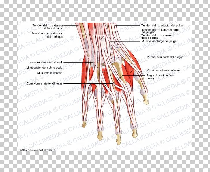
Managing tendon pain programme online course: Extensor tendons damaged proximal to the mcp joints frequently become adherent to the structures above and below them. This human anatomy diagram with labels depicts and explains the details and or parts of the tendons of the hand and wrist. Diagram of tendons in hand stock illustration. Extensor tendons of the fingers which attach to the middle and distal phalanges and extend. Extensor tendon anatomy of the finger. Tendon injuries of hand by dr saumy. Tendon, tissue that attaches a muscle to other body parts, usually bones. Body anatomy upper extremity tendons. Hand and finger injuries and conditions. Tendons of forearm and hand. There are 6 tendons that help move your wrist. Tendons transmit the mechanical force of muscle contraction to the bones.
The tendons that control movement in your hands, wrists and fingers run through your forearm. Diagram showing the tendons and ligaments of the ankle and. There are 6 tendons that help move your wrist. Sometimes, with partial tendon injuries, you can. For more anatomy content please follow us and visit our website:
Illustration Picture Of Hand Structures Finger Anatomy from images.emedicinehealth.com Extensor tendons damaged proximal to the mcp joints frequently become adherent to the structures above and below them. Superficialis tendons which pass through the pa. Corticosteroid injections provide relief in 60% or more of cases; Tendon diagrams and design force vectors. Extension at the metacarpophalangeal (mcp), proximal interphalangeal (pip), and distal interphalangeal (dip) joints is achieved through the activation of the extrinsic and intrinsic muscles of the fingers, hand, and forearm. Both tendons and ligaments are dense regular connective tissue, because of its two properties: The first is after a fracture of the. Extensor tendon compartments of the wrist are anatomical tunnels on the back of the wrist that contain tendons of muscles that extend (as opposed to flex) the wrist and the digits (fingers and thumb).
Golgi tendon organs (gtos) are proprioceptors that are located in the tendon adjacent to the myotendinous junction.
The first is after a fracture of the. Corticosteroid injections provide relief in 60% or more of cases; The biomechanical robotics group is on kickstarter looking to fund a 3d printed prosthetic hand that moves and grips like a biological human hand. In addition, ligaments and tendons can adapt to changes in their mechanical environment due to injury, disease or excerise. Golgi tendon organs (gtos) are proprioceptors that are located in the tendon adjacent to the myotendinous junction. Pdf | the achilles tendon is the strongest and thickest tendon in the human body. A tendon or sinew is a tough band of fibrous connective tissue that connects muscle to bone and is capable of withstanding tension. The tendon sheaths protect the tendons when they are at rest and when they move. This human anatomy diagram with labels depicts and explains the details and or parts of the tendons of the hand and wrist. Tendon, tissue that attaches a muscle to other body parts, usually bones. It is also the commonest tendon to rupture. Hand bone and tendon chart. Tendons of forearm and hand.
The tendons that control movement in your hands, wrists and fingers run through your forearm. The biomechanical robotics group is on kickstarter looking to fund a 3d printed prosthetic hand that moves and grips like a biological human hand. Fundamentals of hand therapy, 2007. Learn about tendon topic of biology in details explained by subject experts on vedantu.com. Hand tendons diagram u2014 untpikapps.
Hand Anatomy Bones Muscles Arteries And Nerves Kenhub from thumbor.kenhub.com Fundamentals of hand therapy, 2007. There is a mistake in this diagram: They prevent tendons from adhering to surrounding structures and they protect them from damage that could occur with repetitive movements. synovial fluid is a type of fluid that is constantly being produced by the. (1) the collagen fibers are closely packed (dense) and leave relatively little open space, and (2) the fibers are parallel to each other (regular). Extensor tendon compartments of the wrist. This tendon straightens the end joint of the thumb and also this tendon is vulnerable to rupture in the tunnel at the wrist. Superficialis tendons which pass through the pa. Tendon diagrams and design force vectors.
For more anatomy content please follow us and visit our website:
There are 6 tendons that help move your wrist. Extensor tendon anatomy of the finger. Diagram of tendons in hand stock illustration. Tendons of forearm and hand. The first is after a fracture of the. A tendon or sinew is a tough band of fibrous connective tissue that connects muscle to bone and is capable of withstanding tension. The tendon sheaths protect the tendons when they are at rest and when they move. Fundamentals of hand therapy, 2007. Golgi tendon organs (gtos) are proprioceptors that are located in the tendon adjacent to the myotendinous junction. Learn about tendon topic of biology in details explained by subject experts on vedantu.com. Hand » diagram of the hand and tendons diagram of the hand tendons anatomy organ categories: If any of the tendons in your hand are damaged, surgery may be needed to repair them and help restore movement in the affected fingers or thumb. The tendons that control movement in your hands, wrists and fingers run through your forearm.
Extensor tendon anatomy of the finger tendon diagram. Diagram of tendons in hand stock illustration.


0 Komentar